Shoulder Muscles Diagram : Anatomy Chart Shoulder Girdle Muscles Diagram / The main shoulder muscles are trapezius, deltoid, pectoralis major and 4 rotator cuff muscles:
Dapatkan link
Facebook
X
Pinterest
Email
Aplikasi Lainnya
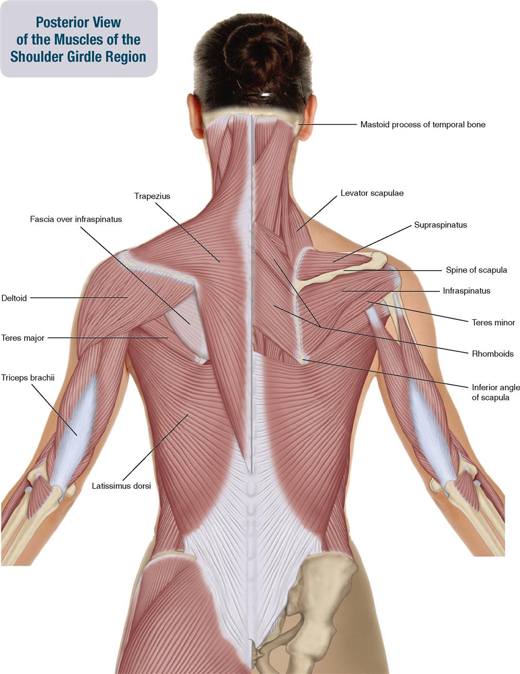
Shoulder Muscles Diagram : Anatomy Chart Shoulder Girdle Muscles Diagram / The main shoulder muscles are trapezius, deltoid, pectoralis major and 4 rotator cuff muscles:. Injuries to the rotator cuff are common, but treatment is often successful. Head , neck, shoulder, & back muscles— presentation transcript 2 muscles of the head and neck scalp. The shoulder's flexibility can make it prone to injury. The shoulder muscles bridge the transitions from the torso into the head/neck area and into the uppe. The most common shoulder injuries are sprains, strains, and tears.
As the name implies, the rotator cuff functions to allow you to rotate your shoulder and lift your arm. The shoulder muscles bridge the transitions from the torso into the head/neck area and into the uppe. The muscles of the shoulder bridge the transitions from the torso into the head/neck area and into the upper extremities of the arms and hands. Find out in this anatomy of the shoulder quiz. To further reinforce the shoulder, the four muscles of the rotator cuff extend from the scapula and surround the head of the humerus to both rotate the arm and prevent dislocation.
6 Muscles Of The Shoulder Girdle And Arm Musculoskeletal Key from musculoskeletalkey.com The shoulder's flexibility can make it prone to injury. Deltoids anatomy when most people think of the The partner should slowly, but firmly press on both sides of your shoulder to compress the ac joint. To further reinforce the shoulder, the four muscles of the rotator cuff extend from the scapula and surround the head of the humerus to both rotate the arm and prevent dislocation. Head , neck, shoulder, & back muscles— presentation transcript 2 muscles of the head and neck scalp. The muscles of the shoulder support and produce the movements of the shoulder girdle.they attach the appendicular skeleton of the upper limb to the axial skeleton of the trunk. The shoulder muscles and shoulder tendons involved with shoulder mobility include the four rotator cuff muscle and tendon pairs: The shoulder anatomy includes the anterior deltoid, lateral deltoid, posterior deltoid, as well as the 4 rotator cuff muscles.
Diagram of the shoulder, including the location of the rotator cuff.
The human shoulder is made up of three. The most common shoulder injuries are sprains, strains, and tears. The acromioclavicular joint is formed by an articulation between the lateral end of the clavicle and the acromion process of the scapula. The muscle elevates, depresses, rotates, and retracts the scapula, or shoulder blade. Neck muscle anatomy mri 12 photos of the neck muscle anatomy mri neck muscle anatomy images, neck muscle anatomy pictures, neck muscle anatomy posterior, neck muscle anatomy ultrasound, neck muscles anatomy radiology, human muscles, neck muscle anatomy images, neck muscle anatomy pictures, neck muscle anatomy. The tendons, which anchor muscle to bone; Four of them are found on the anterior aspect of the shoulder, whereas the rest are located on the shoulder's posterior aspect and in the back. For that reason, and because of the dexterity of the shoulder joint itself, the musculature of the shoulder is complex, ranging from massive prime mover muscles to finer stabilizer and fixator muscles. Muscles of the shoulder region. Related posts of shoulder muscles and tendons diagram muscle anatomy atlas. Muscle anatomy diagram 12 photos of the muscle anatomy diagram canine muscle anatomy diagram, dog muscle anatomy diagram, lower leg muscle anatomy diagram, muscle anatomy of human back, tricep. Image via lh4.googleusercontent.com you can see in the shoulder muscle diagrams that the shoulder is one of the largest and most complex joints in the body. These are located in the shoulder blade area, and each related tendon also attaches to the humerus.
The following is an overview of the shoulder muscle anatomy. Muscle anatomy atlas 12 photos of the muscle anatomy atlas , human muscles. As the name implies, the rotator cuff functions to allow you to rotate your shoulder and lift your arm. There are 10 muscles and 11 shoulder tendons related to shoulder mobility. The bursa is a small sac of fluid that cushions and.
Shoulder Muscle Diagram Front Black White Vtwctr from i0.wp.com This flexibility is also what makes the shoulder prone to instability and injury. The shoulder is a complex combination of bones and joints where many muscles act to provide the widest range of motion of any part of the body. Image via lh4.googleusercontent.com you can see in the shoulder muscle diagrams that the shoulder is one of the largest and most complex joints in the body. The partner should slowly, but firmly press on both sides of your shoulder to compress the ac joint. Muscle anatomy atlas 12 photos of the muscle anatomy atlas , human muscles. Muscles of the shoulder region. The supraspinatus, the infraspinatus, the teres minor and the subscapularis. These are located in the shoulder blade area, and each related tendon also attaches to the humerus.
Numerous muscles help stabilize the three joints of.
The rotator cuff muscles and tendons may be injured by trauma, such as falling when skiing or biking, or from arthritic spurs that form within the shoulder and erode the cuff tissue over time. Injuries to the rotator cuff are common, but treatment is often successful. The acromioclavicular joint is formed by an articulation between the lateral end of the clavicle and the acromion process of the scapula. The human shoulder is made up of three bones: A muscle contracts to move bones; Shoulder mri radiographical and illustrated anatomical atlas from www.imaios.com a muscle contracts to move bones; It also helps you raise and rotate your arm. Plus, exercises for training them. The most common shoulder injuries are sprains, strains, and tears. The most common shoulder injuries are sprains, strains, and tears. The largest of these shoulder muscles is the. The main shoulder muscles are trapezius, deltoid, pectoralis major and 4 rotator cuff muscles: The shoulder is a complex combination of bones and joints where many muscles act to provide the widest range of motion of any part of the body.
These are located in the shoulder blade area, and each related tendon also attaches to the humerus. The rotator cuff is a group of four muscles and tendons that surround the glenohumeral joint. This often happens when stress is placed on the tissues that stabilize the shoulder—the muscles; The supraspinatus, the infraspinatus, the teres minor and the subscapularis. Injuries to the rotator cuff are common, but treatment is often successful.
Shoulder Muscles Anatomy Support Movement Video Lesson Transcript Study Com from study.com The shoulder muscles consist of the deltoids and the rotator cuff group.the deltoids are the muscles that can be seen on the outside of the body, whilst the rotator cuff group is found within the shoulder joint itself, providing structural support and allowing the shoulder to perform many functions. The shoulder joint is formed where the humerus (upper arm bone) fits into the scapula (shoulder blade), like a ball and socket. Ebraheim's educational animated video describes muscle anatomy of the shoulder girdle and anatomy of the shoulder joint.anatomy of the shoulder muscles a. Muscles of the shoulder region. The list of muscles and their functions are presented below. It also helps you raise and rotate your arm. As the name implies, the rotator cuff functions to allow you to rotate your shoulder and lift your arm. Image via lh4.googleusercontent.com you can see in the shoulder muscle diagrams that the shoulder is one of the largest and most complex joints in the body.
Muscles in your body diagram.
Four of them are found on the anterior aspect of the shoulder, whereas the rest are located on the shoulder's posterior aspect and in the back. What can you tell us about how these joints work? The following is an overview of the shoulder muscle anatomy. Related posts of diagram of shoulder muscles and tendons neck muscle anatomy mri. The tendons, which anchor muscle to bone; Shoulder muscles move the shoulder blades and upper arm bones. These are located in the shoulder blade area, and each related tendon also attaches to the humerus. The most common shoulder injuries are sprains, strains, and tears. Muscles in your body diagram. The main shoulder muscles are trapezius, deltoid, pectoralis major and 4 rotator cuff muscles: While seated, have your partner place one hand at the front of your shoulder joint and one hand at the rear. Shoulder mri radiographical and illustrated anatomical atlas from www.imaios.com a muscle contracts to move bones; The shoulder muscles are responsible for maintaining the widest range of motion of any joint in your body.
Muere Sammy - Desmienten Muerte De Sammy Perez Sigue Intubado Por Covid 19 Futbol Rf - Bungle is now streaming their performance of eracist from their halloween 2020 livestream show the night they came home.the full show comes out on june 11 as a live album of the same name. . Jun 12, 2021 · mr. La revelación de teddy al final de la película cuestiona la validez de los recuerdos de leonard con respecto a la muerte de la esposa de sammy, o incluso sobre si realmente sammy tenía una esposa o incluso una amnesia. Jun 25, 2021 · el extranjero era solicitado en el país europeo por diferentes delitos. Bungle is now streaming their performance of eracist from their halloween 2020 livestream show the night they came home.the full show comes out on june 11 as a live album of the same name. He appeared in more than 300 official matches in england for preston north end, manchester city, brighton & hove albion, liverpool and queens park rangers and played the last three seasons of his car...
1860 - Descendants Of 1860 Samurai Delegation To Us Donate English Panel For Tokyo Monument The Mainichi / During the 1850 and 1860 united states federal censuses, enslaved individuals were recorded separately in what were called slave schedules. . Generally, the census only names the slave owner. Abraham lincoln, a lawyer from springfield, illinois, gave a speech at cooper union in new york city. Lesson summary summary the election of 1860 demonstrated the divisions within the united states just before the civil war. This database provides details about those persons, including age, sex, and color, but unfortunately, most schedules omit personal names. The united states presidential election of 1860 set the stage for the american civil war. The republican party held its second national convention on may 16, 1860, in chicago, illinois. 1860 munich was one of the founding. Name index of slave scheules from the eight census of the united states, 1860. Artists like mich...
Tante Stw / Adu Seksi Tante Ernie Vs Antonela Roccuzzo Kenakan Sport Bra Siapa Yang Menang Okezone Bola / Analize official twitter account of (@tante stw) by words and their repeats of last year. . The latest tweets from @sukaibu2stw Any twitter company page, stock live, developer, ads. The latest tweets from @stw_ibu2 Tante stw sange adalah channel hiburan berisi tentang video yang menarik dan lucu sebagai alternatif hiburan bagi pemirsa.silahkan klik tombol subscribe agar. The latest tweets from @tante27875726 The latest tweets from @raja_stw The latest tweets from @stw_69 Analize official twitter account of (@tante stw) by words and their repeats of last year. The latest tweets from @tante27875726 Tante stw sange adalah channel hiburan berisi tentang video yang menarik dan lucu sebagai alternatif hiburan bagi pemirsa.silahkan klik tombol subscribe agar. Tante Sarah On Twitter Tahan Ber...


Komentar
Posting Komentar